In summary

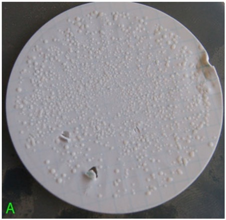
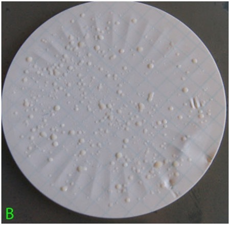
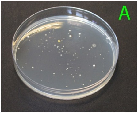
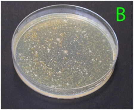
  • Pathogen testing in water is generally performed as presence/absence, due to low pathogen concentrations that defy accurate quantification.
  • Test methods include membrane filtration, most probable number (MPN) determination, and direct plating methods.
  • Test results should be tracked over time using the same lab and method for valid comparison.
  • If indicator results exceed your limits, suggested actions include:
    • maximising the interval between last irrigation and harvest
    • switching water sources
    • changing irrigation methods
    • treating the water before use.

Human pathogens commonly linked with irrigation water are mainly enteric bacteria, which originate in the gut and spread in the environment via livestock and wildlife faeces. Examples include Campylobacter, Salmonella, and Escherichia coli O157. Though not strictly enteric, Listeria monocytogenes, is also significant; it is found ubiquitously in soil, water, and manure (1). Because it thrives in refrigeration temperatures and has a high fatality rate (~35%), it is treated with special caution in post‑harvest hygiene monitoring.

Laboratory test methods for water samples

Water is tested in the laboratory by:

A.    Membrane filtration, concentrating bacteria on a membrane then culturing, shown in Figure 2 (before/after UV treatment).
B.    Most Probable Number (MPN), which uses serial dilutions and statistical tables, useful for low density or turbid samples.
C.    Direct plating, where samples are either spread or mixed with agar. Colony results are illustrated in Figure 3B after incubation.

A. Membrane Filtration

The filter-based method involves passing a 100 ml water sample through a membrane filter with pores that trap bacteria while allowing water to pass through. This concentrates any bacteria present on the surface of the membrane. The membrane is then placed on a solid nutrient medium, allowing individual bacterial cells to grow into visible colonies that can be counted. Figure 1 shows typical results from this method, illustrating the appearance of cultured membranes from a fast-flowing river both before and after ultraviolet (UV) light treatment.

Figure 1: A set of typical incubated water filter membranes from a fast-flowing overland river before treatment (A)

A set of typical incubated water filter membranes from a fast-flowing overland river before treatment

A set of typical incubated water filter membranes from a fast-flowing overland river before after treatment (B)

A set of typical incubated water filter membranes from a fast-flowing overland river after treatment (B)

B. Most Probable Number (MPN) Method

The most probable number (MPN) method, although often considered more traditional, remains valuable. It is particularly useful when the water contains high levels of dissolved solids that may clog filtration membranes, or when bacterial levels are extremely low, below the detection threshold of other methods such as direct plating. Unlike the filter method, MPN provides a statistical estimate of bacterial concentration rather than a direct count.

In an MPN test, small sample volumes are serially diluted (typically at factors of 10, 100, 1,000, 10,000 and 100,000), and a fixed number of volumes (usually 3 or 5) from each dilution are added to tubes of liquid growth medium. After incubation, the number of tubes showing bacterial growth at each dilution is recorded and cross-referenced against established tables to estimate the most probable bacterial concentration in the original sample. Laboratories usually report MPN results as MPN/100 ml, distinguishing it from filter methods, which are reported as cfu/100 ml.

C. Direct Plating Method

Direct plating is a straightforward technique often used to count total mesophilic aerobic bacteria, microbes that thrive at moderate temperatures and require oxygen. In this method, small volumes of either undiluted or diluted water are spread directly onto the surface of an agar plate or mixed into warmed, liquid agar before solidification. After incubation, visible colonies form from individual bacterial cells, allowing for enumeration. Figure 2 illustrates results from a mesophilic aerobic count test using two different dilutions of the same sample.

Figure 2: (A) Petri dish showing the range of different bacterial colonies after incubation of a highly diluted sample (1:1,000,000)

Petri dish showing the range of different bacterial colonies after incubation of a highly diluted sample (1:1,000,000).

Figure 2: (B) A lower dilution of the same water sample (1:1,000), showing too many colonies to count individually

(B) A lower dilution of the same water sample (1:1,000), showing too many colonies to count individually.

Regardless of the method used, results for water samples are generally reported as colony-forming units per 100 millilitres (cfu/100ml or MPN/100 mL), as it is uncommon to detect bacteria in just 1 ml of good-quality water. In comparison, food samples are typically tested using a 25 g portion, and results are reported as cfu/g.

Interpretation of test results

In the UK, there are no legally binding standards for irrigation water quality unless the water is recycled wastewater. Unless growers are attempting compliance with a microbiological standard within an assurance scheme or that required by a specific retailer, growers should choose an indicator standard from international guidance.

An overview of selected international guidance standards is provided (table 1). 

Table 1: International standards and guidelines for selected indicator bacteria numbers in irrigation and potable water. (RTE = ready‑to‑eat; MPN = most probable number, MPN is equivalent to cfu and the use depends on the method used to determine bacterial load, both are valid measures of bacterial density)

Issuing bodyIndicator bacteriaPerformance criteria
World Health Organisation Irrigation water for crops to be eaten rawFaecal coliforms≤1000 cfu/100 ml (calculated as geometric mean)
UK Drinking Water Inspectorate: Service reservoir waterE. coli and also faecal coliforms0 cfu/100 ml for both indicators in 95% of samples 
State of California, USA. Recycled irrigation water for RTWTotal coliforms≤2.2 MPN cfu/100 ml in previous 7 days of test results. No sample to exceed 230 MPN/100 mL in previous month
Canadian Agriculture Ministry Irrigation water guidelinesFaecal coliforms or E. coli and also total coliforms100 cfu/100 ml of faecal coliforms or E. coli; 1000 cfu/100 ml total coliforms 

Testing should be conducted at intervals determined by a chosen standard, such as Red Tractor, or at the grower’s discretion. For those not following a formal scheme, monthly testing is often considered a good starting point. Over time, plotting these results helps build a picture of how water microbiology fluctuates. More information on trending and how to interpret trending data is available.  A typical example of a water quality trend from a river used for irrigation is presented in Figure 3.

Figure 3

Out-of-Specification Results

Routine monitoring combined with reactive intervention, only taking corrective action when results exceed acceptable limits, is generally the most cost-effective strategy for growers seeking to produce safe, microbiologically sound crops. While testing itself does not change the water quality, growers often opt for more frequent testing following an out-of-specification result. This provides a clearer view of changes during periods of concern.

When a result falls outside acceptable parameters, several corrective actions may be considered. The chosen response will depend on available financial resources and the severity of the contamination. First, a thorough risk assessment should be conducted to identify potential contamination sources and inform the best course of action. Options include:

  1. increasing the water interval, i.e. the time between last irrigation and harvest: Pathogens tend to decline rapidly on crop surfaces exposed to sunlight and dry conditions. In hot weather, avoid irrigating shortly before harvest, as this can result in high E. coli levels if the water is contaminated. More information on water intervals here.
  2. switching to an alternative water source: Often the simplest option is to change to a supply that meets the specification. Though costly, mains water is the highest quality option. However, blending out-of-spec water with mains water is discouraged, as it does not address the underlying contamination source. More information on water sources here.
  3. changing the irrigation method: The risk from contaminated water increases significantly when the water is in contact with the edible portion of the crop. Switching to irrigation systems like drip or trickle tape, which avoid direct contact with the produce can mitigate this risk when lower quality water must be used. More information on water contact here.
  4. treating the water before use: Although expensive, treatment systems such as ultraviolet (UV) light units or reverse osmosis filters can effectively reduce microbial loads. Chemical treatments (e.g. ozone or hypochlorite-based purifiers) can also provide a cost-effective solution for treating smaller volumes. Where no clean alternative water source exists and other mitigations are impractical, treatment may be the only viable option. More information on water treatment here.

Important considerations for laboratories and data interpretation

When analysing trends in microbiological results over time, consistency in testing is crucial. The same test method must be used consistently, and each laboratory report should include a reference to the method used. This allows for comparison with previous data. Results obtained using different methods, even on the same sample, can vary considerably. Similarly, different laboratories using the same method may produce significantly different results due to variations in equipment, growth media, and procedural nuances. It is considered poor practice to plot test results from different laboratories on the same trend graph. If a new laboratory or method is introduced, a new graph should be started to avoid misinterpretation.

High-quality laboratories also define and report their detection limits. Rather than reporting low counts as zero, they will indicate values below the detection limit with a ‘less than’ symbol. For example, if the detection limit is 10 cfu/100 ml and no bacteria are detected, the result will be reported as <10 cfu/100 ml. When plotting or calculating averages (such as geometric means), it is common practice to use half the detection limit for such results, so <10 cfu/100 ml would be recorded and analysed as 5 cfu/100 ml.

Did you find this helpful? We would love to hear from you.
Thank you for your feedback!